News
Bảng tin
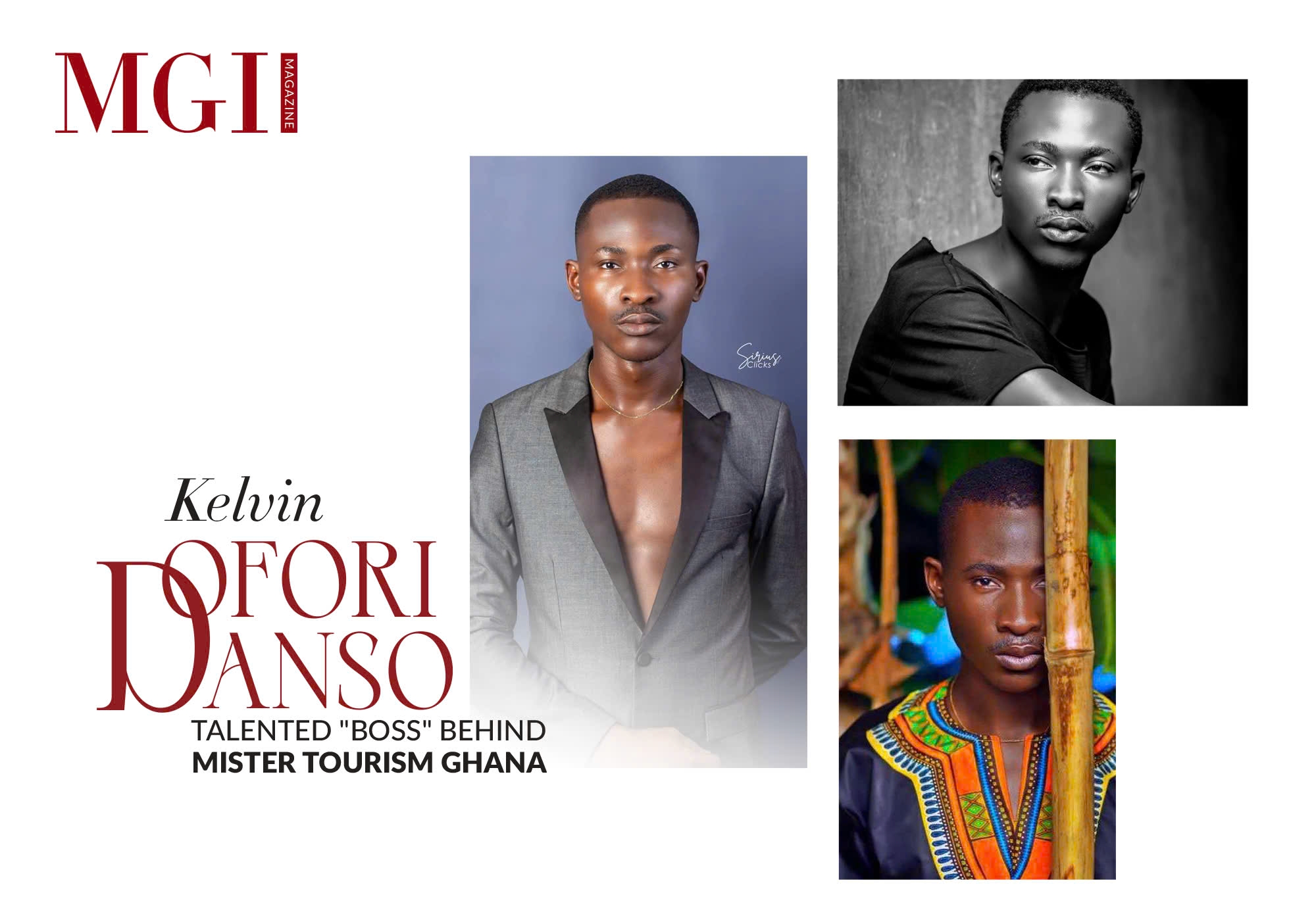
Kelvin Ofori Danso - Talented "boss" behind Mister Tourism Ghana
Mr. Kelvin Ofori Danso, also known as Kelly Ofori Tailormodel, is the National Director of the Mister Tourism Ghana pageant. Coming from a modeling background, Mr. Kelvin Ofori Danso has gained extensive experience on numerous prestigious fashion runways and has held important positions in the fashion industry in Ghana. In addition, this talented director has achieved remarkable success in various other fields.
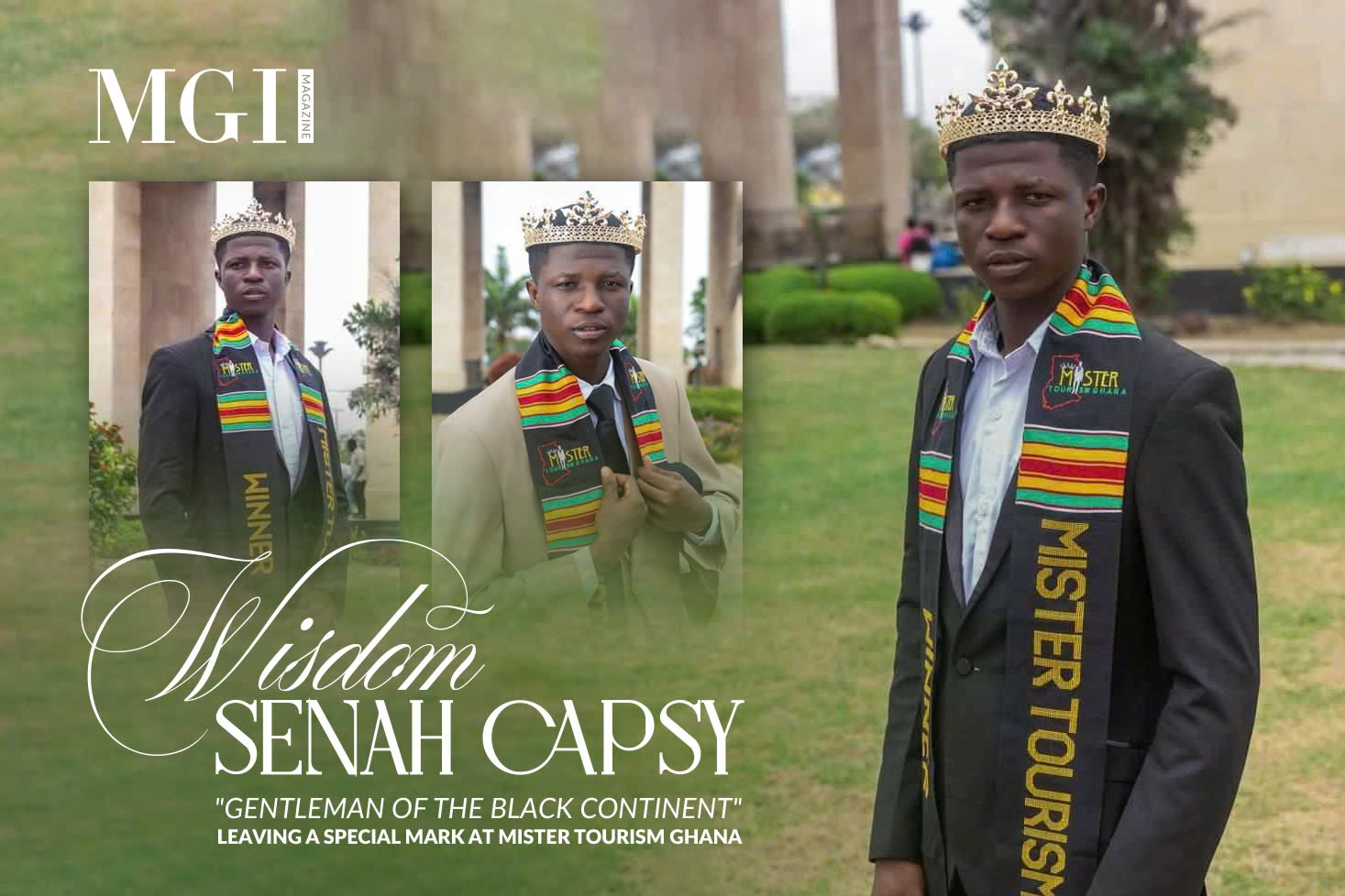
Wisdom Senah Capsy - "Gentleman of the Black Continent" leaving a special mark at Mister Tourism Ghana
In the search for a gentleman with influence, who cares about environmental issues, nature conservation, tourism development, and culture in Ghana, Mister Tourism Ghana was created to find a deserving gentleman. Recently, Wisdom Senah Capsy excelled and surpassed the strong contenders to officially win the title, leaving a new mark at Mister Tourism Ghana 2024, marking a completely new milestone in his journey of pursuing his passion.
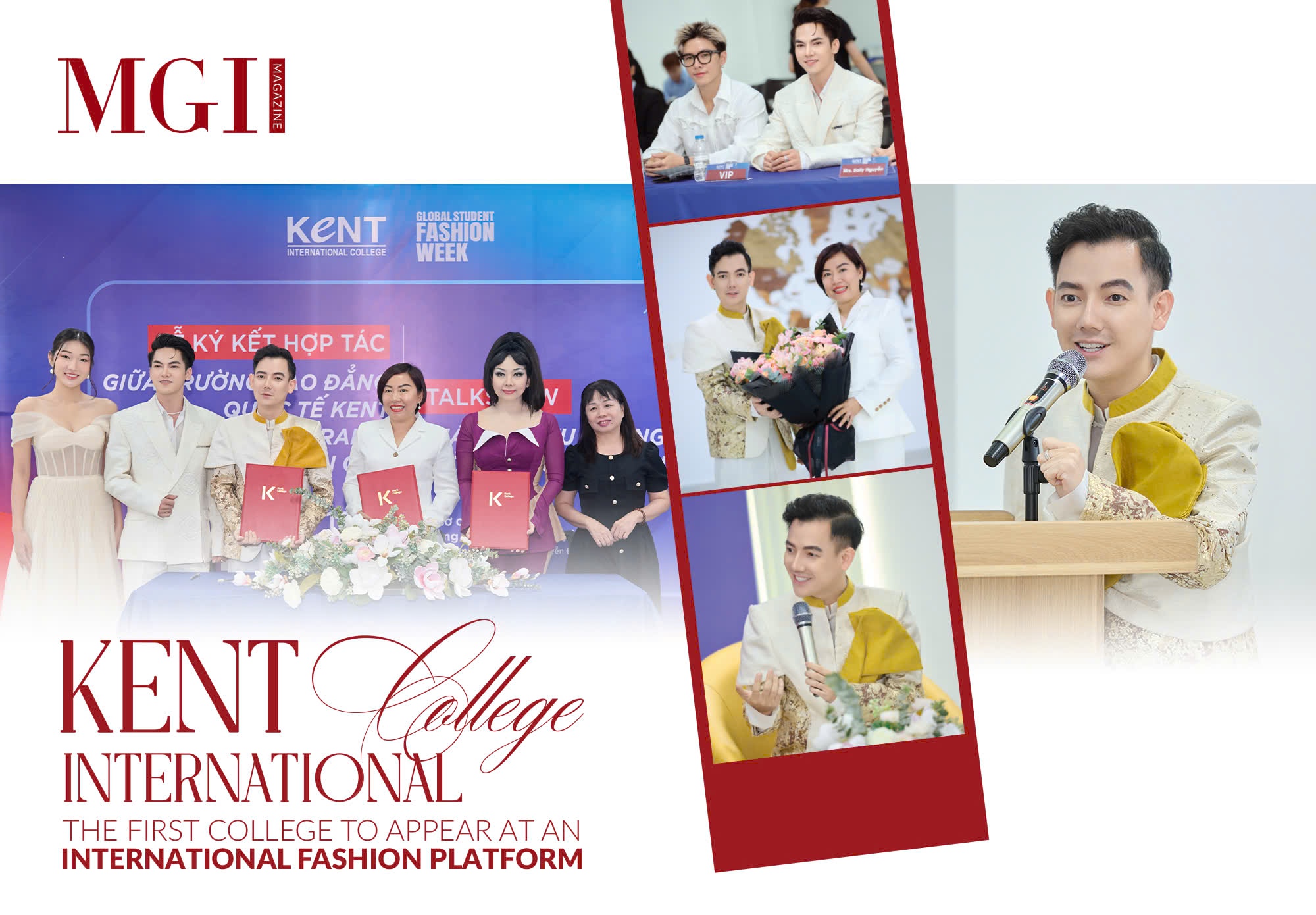
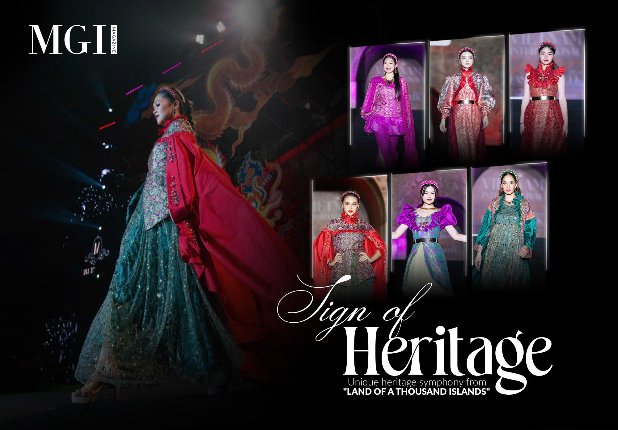
KENT International College - The first college to appear at an international fashion platform
Global Student Fashion Week (GSFW) is the world’s first fashion week dedicated to educational institutions in the fashion field, with students as its primary focus. With a mission to create a new era for the global fashion industry, the inaugural Global Student Fashion Week in 2025 promises to deliver truly spectacular performances that will leave you amazed. As one of 30 prestigious fashion schools, Kent International College has officially signed on with Global Student Fashion Week, becoming the first college to participate in this international fashion arena.
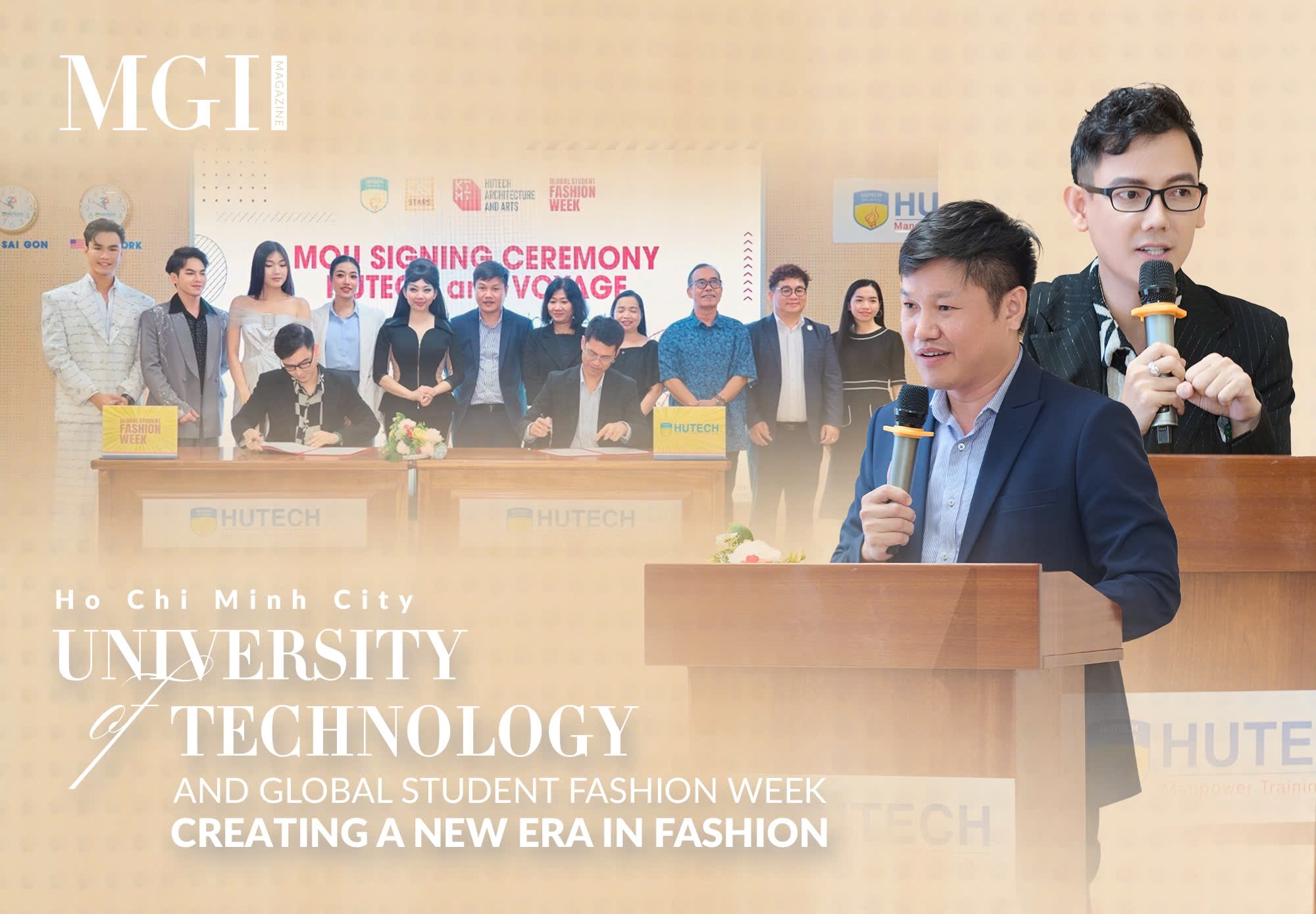
Ho Chi Minh City University of Technology and Global Student Fashion Week creating a new era in fashion
On the morning of August 7, 2024, the signing ceremony of the cooperation between Ho Chi Minh City University of Technology (HUTECH) and the Global Student Fashion Week officially took place. The ceremony was attended by representatives of the university and Mr. Le Tran Dac Ngoc – Chairman of Global Student Fashion Week.
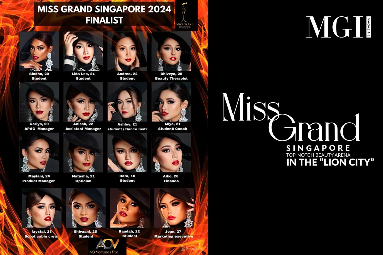
Miss Grand Singapore - Top-notch beauty arena in the “Lion City”
The race for the prestigious crown is heating up. The contestants in Singapore are continually striving to prove themselves and become the most deserving representatives for Miss Grand International 2024. Will any of the beauties surpass Jiahui Jade Wu—Miss Grand Singapore 2023 and the holder of the Grand Voice title at Miss Grand International—to continue competing in this arena?

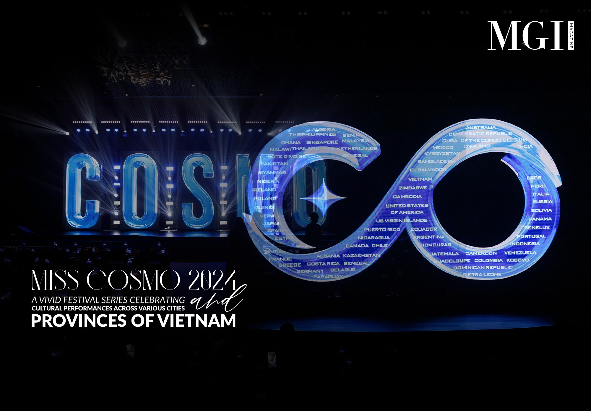
Who will represent Singapore to compete at Miss Cosmo in Vietnam this October?
Miss Cosmo 2024, featuring participants from over 70 countries and territories around the world, will have representatives from these nations officially competing in Vietnam. Through a series of activities during the competition, these representatives will help promote the image of Vietnam’s country, people, and tourism. Additionally, the Miss Cosmo platform offers an opportunity to showcase and celebrate the unique identity of each nation globally. In Singapore, with 12 beautiful contestants, who will become the representative to compete in Miss Cosmo in Vietnam this October?

Mr. Le Tran Dac Ngoc accepts the invitation to judge Miss Grand Singapore 2024
Mr. Le Tran Dac Ngoc - a powerful figure of Global Student Fashion Week, one of the most prestigious fashion shows for global students - has officially accepted the invitation to become a member of the judging panel for Miss Grand Singapore 2024. The unexpected appearance of the GSFW Chairman promises to bring fresh and unique perspectives to this year's prestigious beauty pageant in the “Lion City”.
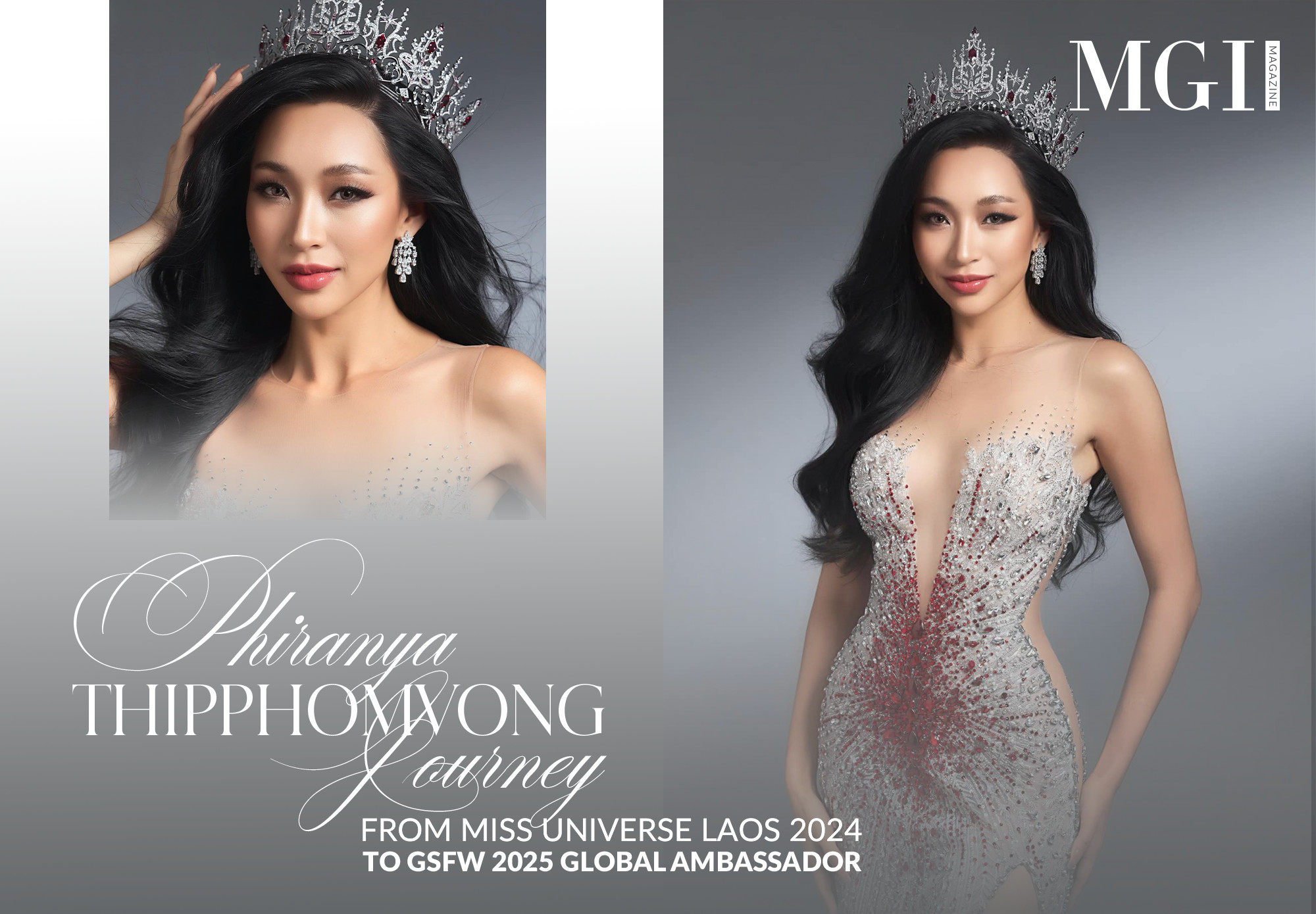
Phiranya Thipphomvong - Journey from Miss Universe Laos 2024 to Global Student Fashion Week 2025 Global Ambassador
Not just a fashion week, Global Student Fashion Week is a journey of connection, empowering young talents to shine on the world fashion map. With the mission of creating a new era for the global fashion industry, Global Student Fashion Week promises to deliver the most sublime and spectacular performances. Kicking off this special journey is the “999 Global Ambassadors” project, featuring the presence of Miss Universe Laos 2024 - Phiranya Thipphomvong.
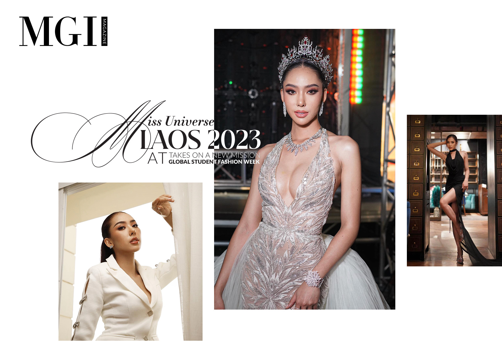
Miss Universe Laos 2023 takes on a new mission at Global Student Fashion Week
Global Student Fashion Week (GSFW) is the world's first fashion week dedicated to students, gathering 999 Global Ambassadors, including Miss Universe titleholders, Mr. Global winners, and influential figures from around the globe. Joining this journey to shape a new era of fashion is Miss Universe Laos 2023, Papao Phaimany Lathsabanthao, in her role as a Global Ambassador, spreading the messages and inspiring the youth.

Miss Queen Laos 2020 accompanying Global Student Fashion Week 2025 as Global Ambassador
The fashion industry has been solidifying its strong position in the modern economy, becoming one of the most attractive fields for young people passionate about creativity. Recognizing the enormous development potential and the rapid changes in market trends, Global Student Fashion Week has officially launched with the aim of creating a dedicated platform for students, allowing the next generation of global designers to express and assert themselves. Joining GSFW 2025 in its journey to promote the global fashion industry is the presence of Ms. Aliya Sirisopha - Miss Queen Laos 2020.